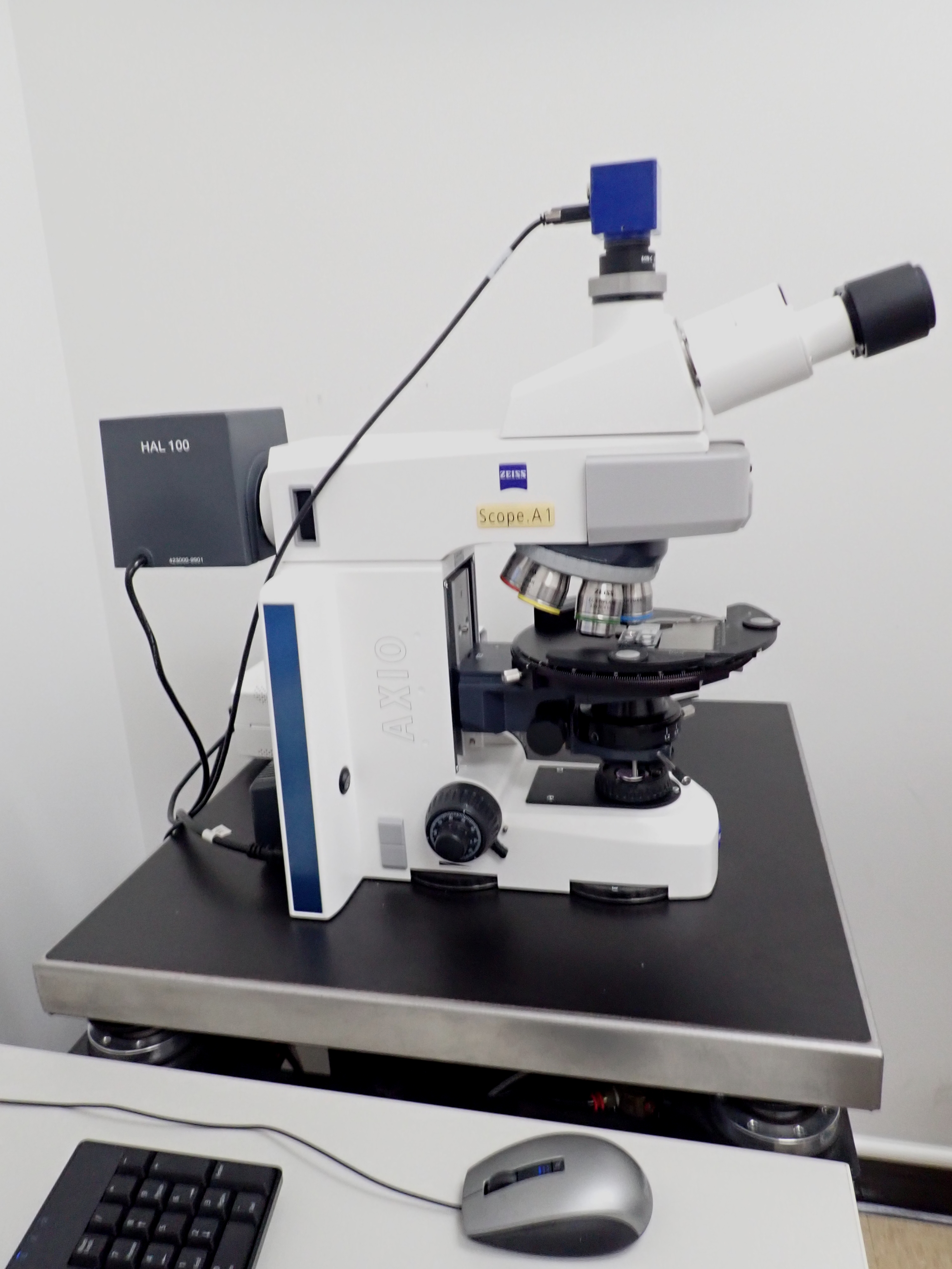
Optical Microscopy

The optical microscope, also referred to as a light microscope, is a type of microscope that commonly uses visible light and a system of lenses to generate magnified images of small objects. Optical microscopes are the oldest design of microscope and were possibly invented in their present compound form in the 17th century. Basic optical microscopes can be very simple, although many complex designs aim to improve resolution and sample contrast.
The object is placed on a stage and may be directly viewed through one or two eyepieces on the microscope. In high-power microscopes, both eyepieces typically show the same image, but with a stereo microscope, slightly different images are used to create a 3-D effect. A camera is typically used to capture the image (micrograph).
The sample can be lit in a variety of ways. Transparent objects can be lit from below and solid objects can be lit with light coming through (bright field) or around (dark field) the objective lens. Polarised light may be used to determine crystal orientation of metallic objects. Phase-contrast imaging can be used to increase image contrast by highlighting small details of differing refractive index.
A range of objective lenses with different magnification are usually provided mounted on a turret, allowing them to be rotated into place and providing an ability to zoom-in. The maximum magnification power of optical microscopes is typically limited to around 1000x because of the limited resolving power of visible light. The magnification of a compound optical microscope is the product of the magnification of the eyepiece (say 10x) and the objective lens (say 100x), to give a total magnification of 1,000×. Modified environments such as the use of oil or ultraviolet light can increase the magnification.
APPLICATIONS
- Use
- Strengths
LIMITATIONS
- Limitations
HIROX KH-7700: 3D Digital Video Microscope
The HIROX KH-7700 3D Digital Video Microscope is an optical inspection microscope used to image objects with rough surfaces or irregular topology, do optical comparisons, measure feature sizes in 2 or 3 dimensions, generate 3D profiles, and view objects from multiple perspectives. The optics are optimized for digital imaging, and it has a significantly larger depth of field than conventional optical microscopes. It also has a motor driven prism system that makes it possible to record streaming video movies of objects viewed from a rotating perspective.
HIROX KH-7700 3D Digital Video Microscope is equipped with:
- 50X-400X zoom lenses for static and rotational viewing at oblique angles
- Magnifications from 1X to 7000X (field of views from 340mm to 0.049mm)
- Diffuse, variable angle, and co-axial lighting; removable fiber optic adapter for flexible lighting arrangements
- Automatic Z-axis control and focusing with software for 3D profilometry
- Software for optical comparison of images with a master pattern
- Static mages recorded with up to 4800×6400 pixel resolution (30 megapixels) in jpg, bmp, or tiff file formats; dynamic image recording in avi format (selectable resolution)
- Portable LCD display unit and lens heads
- DVD multi drive for storing data in DVD-RAM, DVD+/-RW, DVD+/-R, CD-RW or CD-R formats
TECHNICAL SPECIFICATIONS
- Technical
Publications with NanoEarth that acknowledge NSF.
Training modules, videos, sample prep, etc.


