Facilities

NanoEarth is housed primarily in two facilities at the Virginia Tech Institute for Critical Technology and Applied Science (ICTAS). Additionally, NanoEarth has established a partnership with EMSL at PNNL and ENAL at the University of South Carolina to direct users whose specific needs may be better met by their state of the art facilities.
Please note that external users are required to prepare a Purchase Order from their home organization. For cost information, please see the NCFL rates.
Jump to information about facilities at: NCFL // Kelly Hall // EMSL// ENAL.
Check out our User Portal to receive recommendations on which instrument or technique might be able to help you answer your research questions.
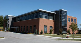
The 15,000 sq. ft. Nanoscale Characterization and Fabrication Laboratory (NCFL) houses a broad array of state-of-the-art electron- and ion-beam characterization tools, as well as office space for staff and visitors.
The NCFL was created to provide researchers with the tools to work in converging disciplines at these dimensions. Established in 2007, it is an initiative of the Institute for Critical Technology and Applied Science at Virginia Tech. The facility is equipped with more than $13 million in highly specialized equipment, more than half of which was made possible through funding provided by Commonwealth Research Initiative. It seeks to help researchers investigate novel phenomena and build transforming technologies that solve critical challenges.
VT NCFL provides:
- Access to advanced equipment for electron microscopy, optical microscopy, and several spectroscopic techniques
- Training for students and researchers in the use of the lab's instrumentation
- Short courses and characterization services for industry.
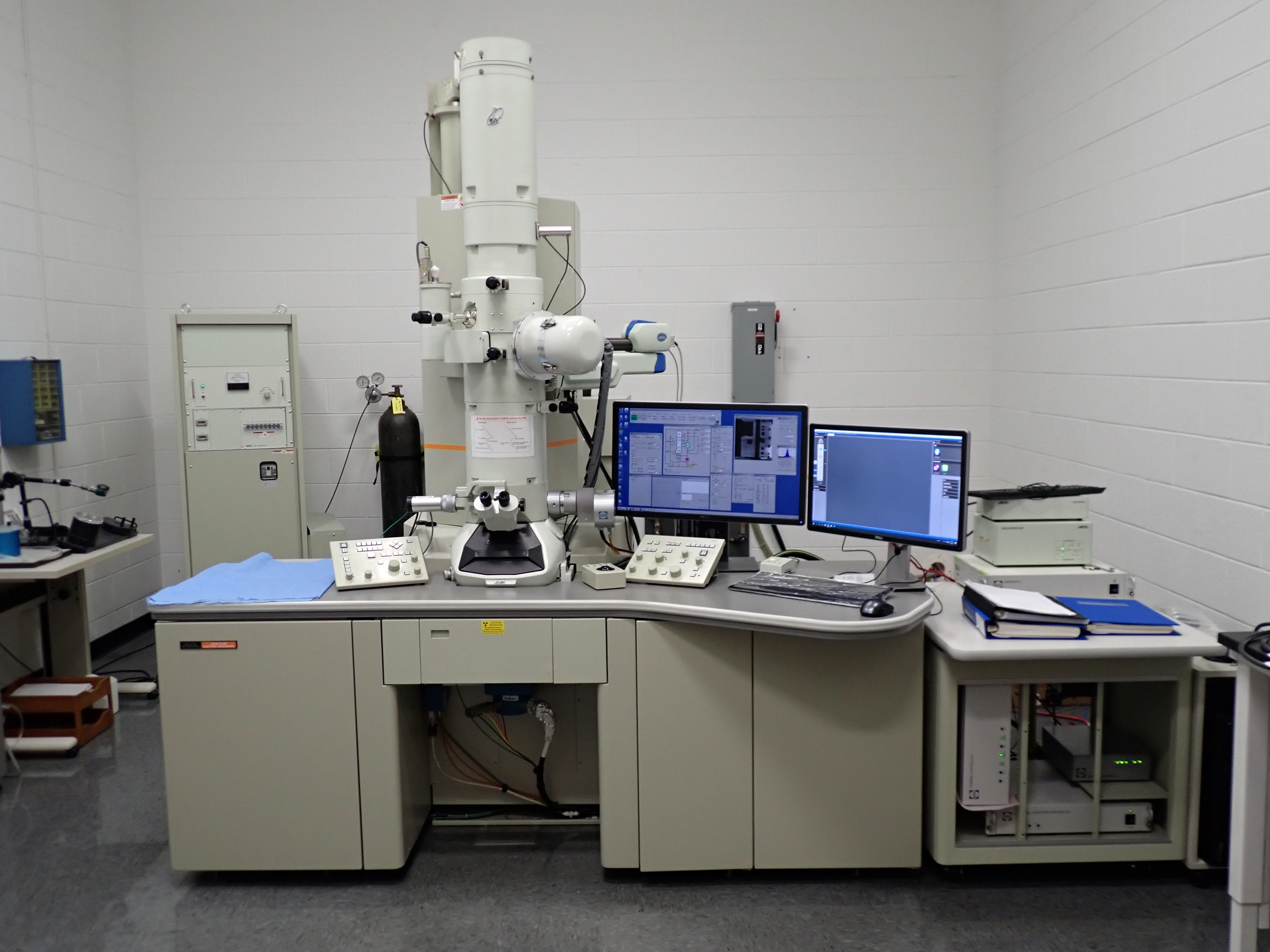
JEOL 2100
A state of the art field thermionic emission analytical electron microscope. The high-resolution pole piece provides 0.2 nm point-to-point resolution and 0.14 nm lattice resolution, enabling high-resolution (HRTEM) imaging. A silicon drift detector-based Energy-dispersive X-ray Spectrometry (EDS) system allows rapid, high-throughput chemical mapping of samples. Typical applications for this instrument are the imaging of soft and hard materials, diffraction analysis of crystalline materials, room temperature tomography, chemical mapping, and general research.
Philips EM420
A conventional TEM, enabled with bright field and dark field imaging and electron diffraction. The microscope is capable of low acceleration voltage operation (from 60 up to 120kV), thus it is ideal for polymer, biological and other electron beam sensitive materials.
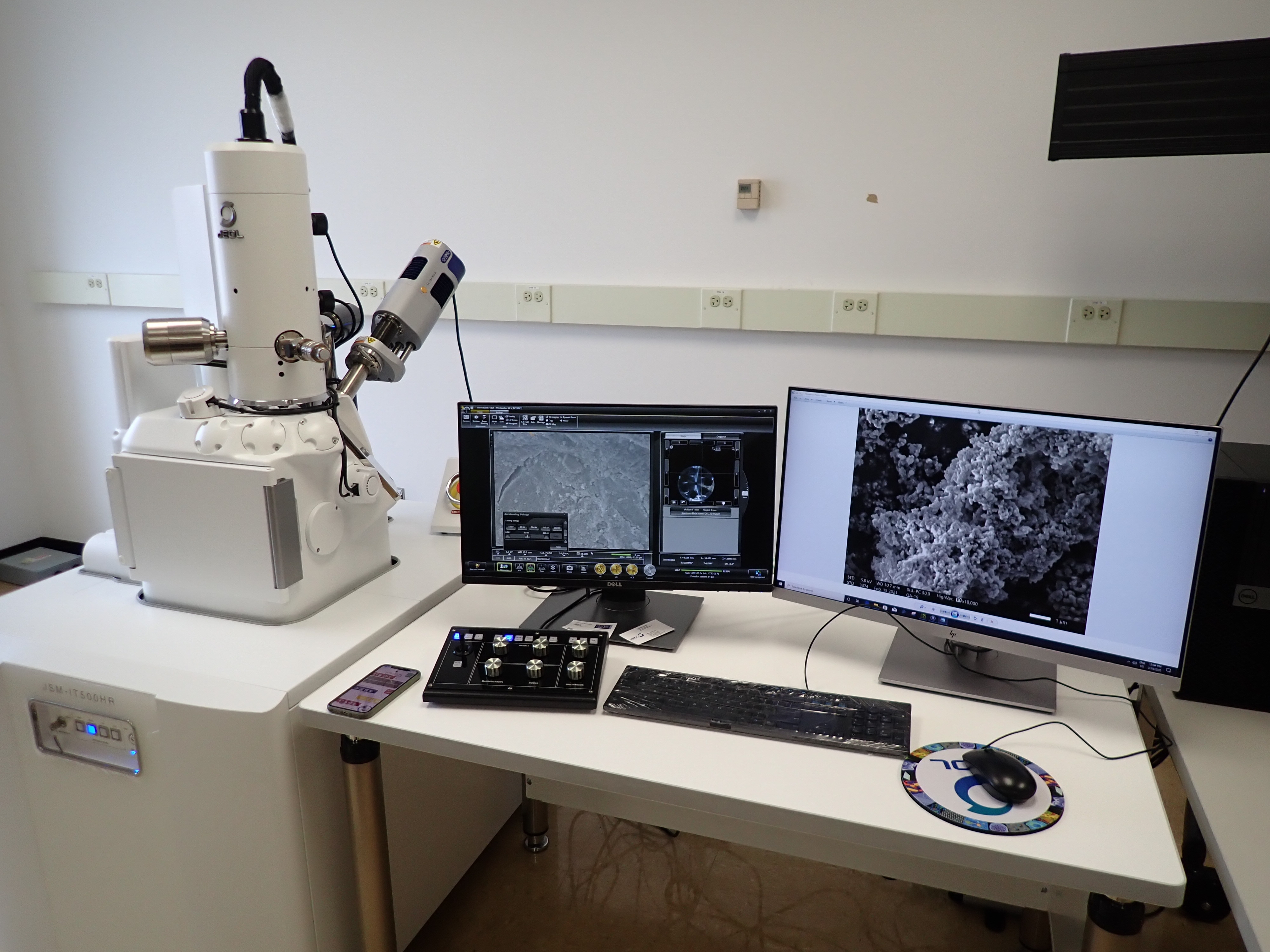
JEOL IT-500 SEM
An analytical FEG SEM offering a seamless link from optical to high magnification SEM images and automated large area imaging and spectroscopy analyses. The IT-500 is also equipped with an Oxford Instruments Ultim® Max EDS silicon drift detector with a 100 mm2 sensor size, allowing for elemental analysis of samples.
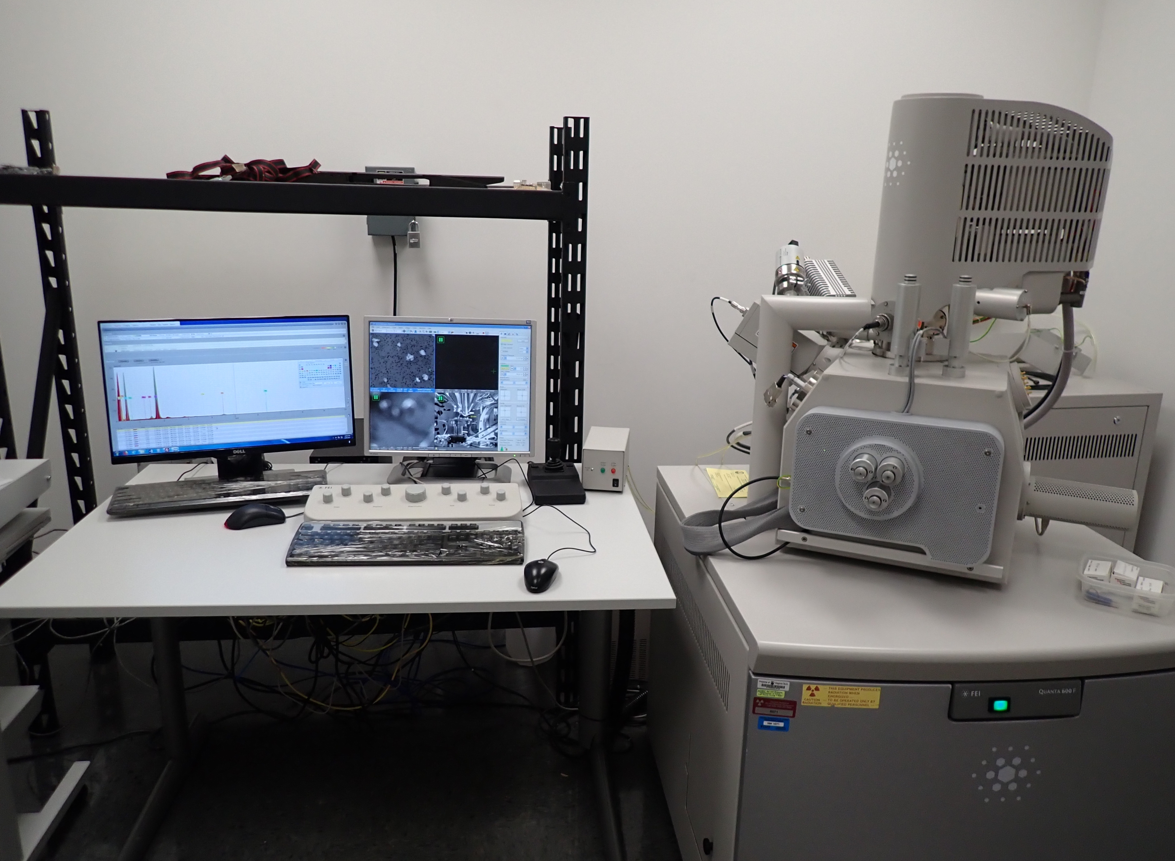
FEI Quanta 600 FEG Environmental SEM
Can operate in high-vacuum and low-vacuum modes. It is used to image samples that are difficult to impossible to image in high vacuum SEMs; in situ experiments such as hydrating, dehydrating and heating samples are possible with the ESEM. It can operate with pressures around the sample up to 4000 Pa and in conjunction with a Peltier stage can image fully hydrated samples, a critical advantage for imaging biological samples. It is equiped with a Bruker Energy-dispersive X-ray Spectrometer (EDS) with a Silicon Drifted Detector.
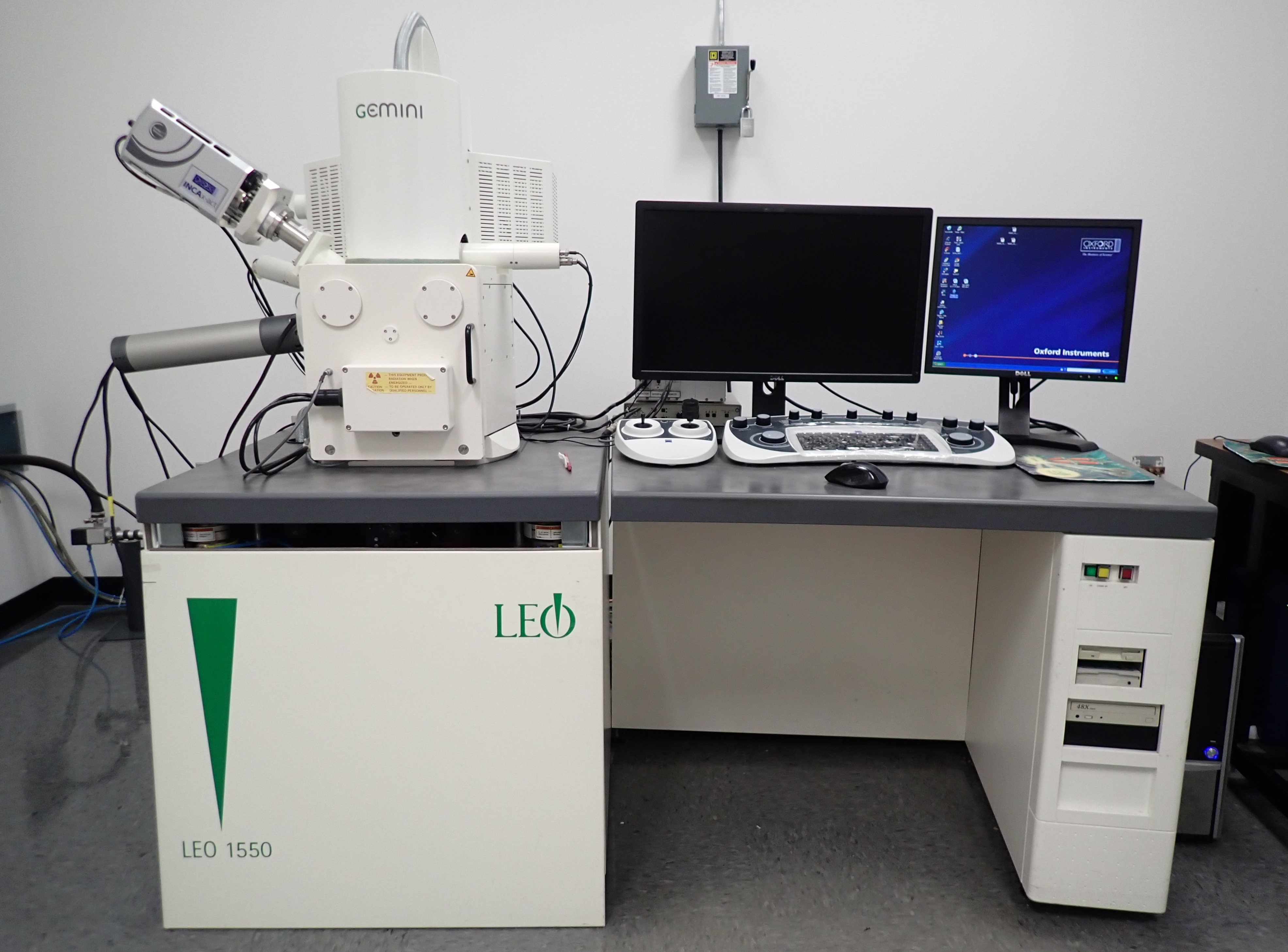
LEO (Zeiss) Field Emission SEM
Capable of resolution in 2-5 nm size range. It is used for high-resolution imaging of surfaces, qualitative assessment of the distribution of elements (with atomic numbers between boron and uranium), submicron structure analysis, and determination of crystal orientation and crystalline texture.
Bruker AFM Multimode, a conventional AFM.
HIROX KH-7700 3D Digital Video Microscope
An optical inspection microscope used to image objects with rough surfaces or irregular topology, do optical comparisons, measure feature sizes in 2 or 3 dimensions, generate 3D profiles, and view objects from multiple perspectives. The optics are optimized for digital imaging and it has a significantly larger depth of field than conventional optical microscopes. It also has a motor driven prism system that makes it possible to record streaming video movies of objects viewed from a rotating perspective.
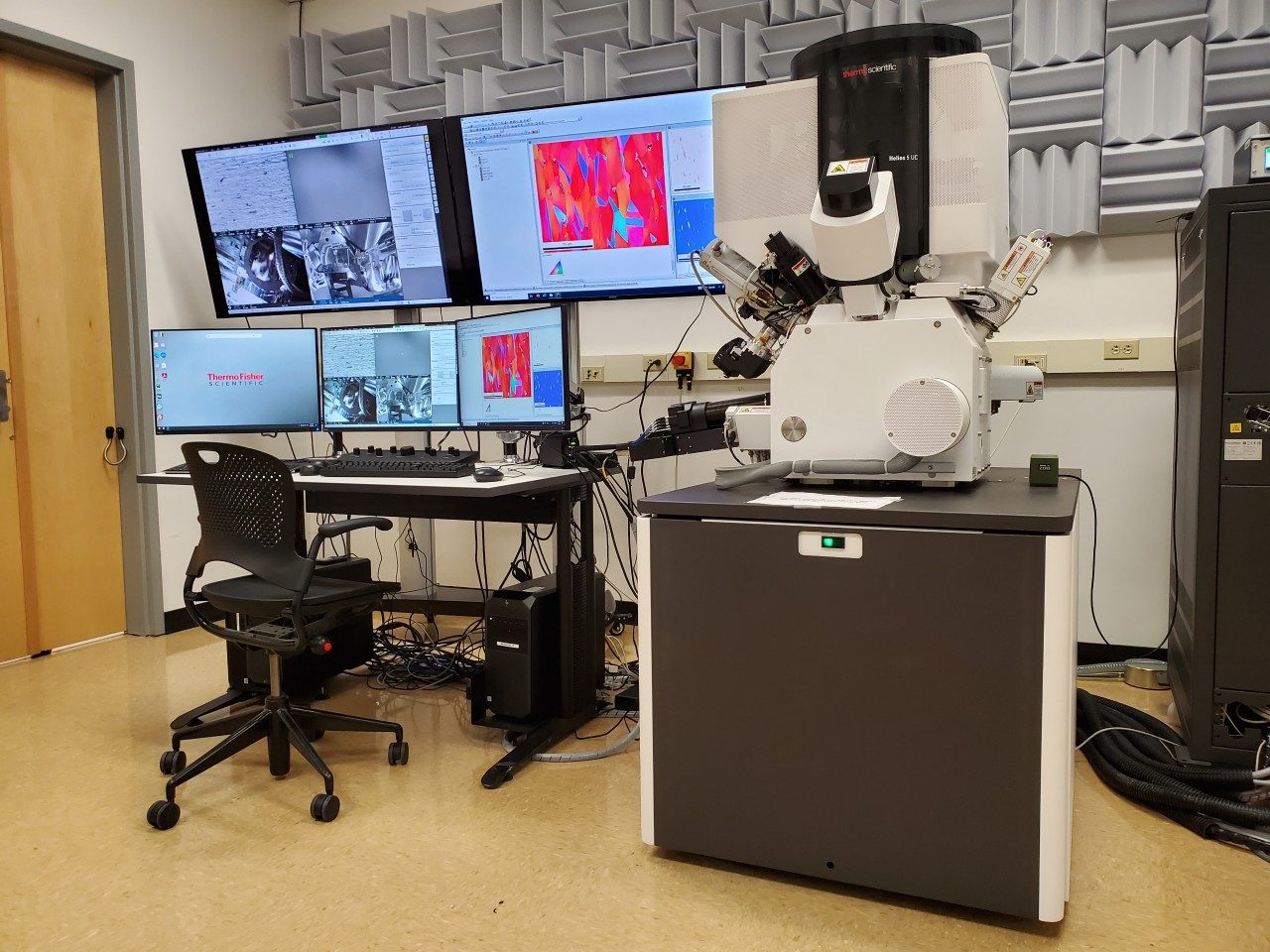
Thermofisher Helios 5 DualBeam Focused Ion Beam (FIB)
A dual-beam workstation that combines a high-resolution SEM and a focused ion beam. The instrument is also equipped with an EDAX electron backscatter detector (EBSD) for crystallographic orientation determination and an EDAX energy dispersive spectrometer (EDX) system for chemical analysis. Thermofisher Scientific AutoTEM 5 software allows for either fully automated or assisted in-situ FIB lift out acquisition, reducing time spent fabricating electron transparent samples.
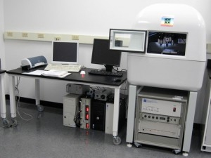
Hysitron TriboIndenter
An automated nanomechanical test instrument for measuring the hardness and elastic modulus of thin films and coatings using controlled indentation of surfaces.
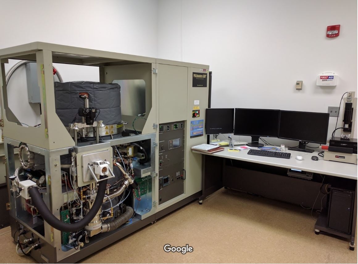
XPS: PHI Quantera SXM
A Scanning Photoelectron Spectrometer Microprobe (XPS or ESCA) used for quantitative analysis of the chemical elements and chemical states within the top few nanometers of a surface. The Quantera features a focussed, monochromated X-ray source for small-spot analysis, and it is automated for high sample throughput. Depth profiling can be accomplished with automated ion milling.
The NCFL has two sample prep rooms with a variety of sample preparation instrumentation, including:
- Cryo ultramicrotome
- Gold and carbon sputterers
- Leica ion slicer
- Gentle mill
- Fischione ion mill
- Ultrasonic disc cutter
- Dimple grinder
- Isomet diamond saw
- Chemical fume hoods
For a full list of instrument capabilities and cost information, please go to the NCFL website.
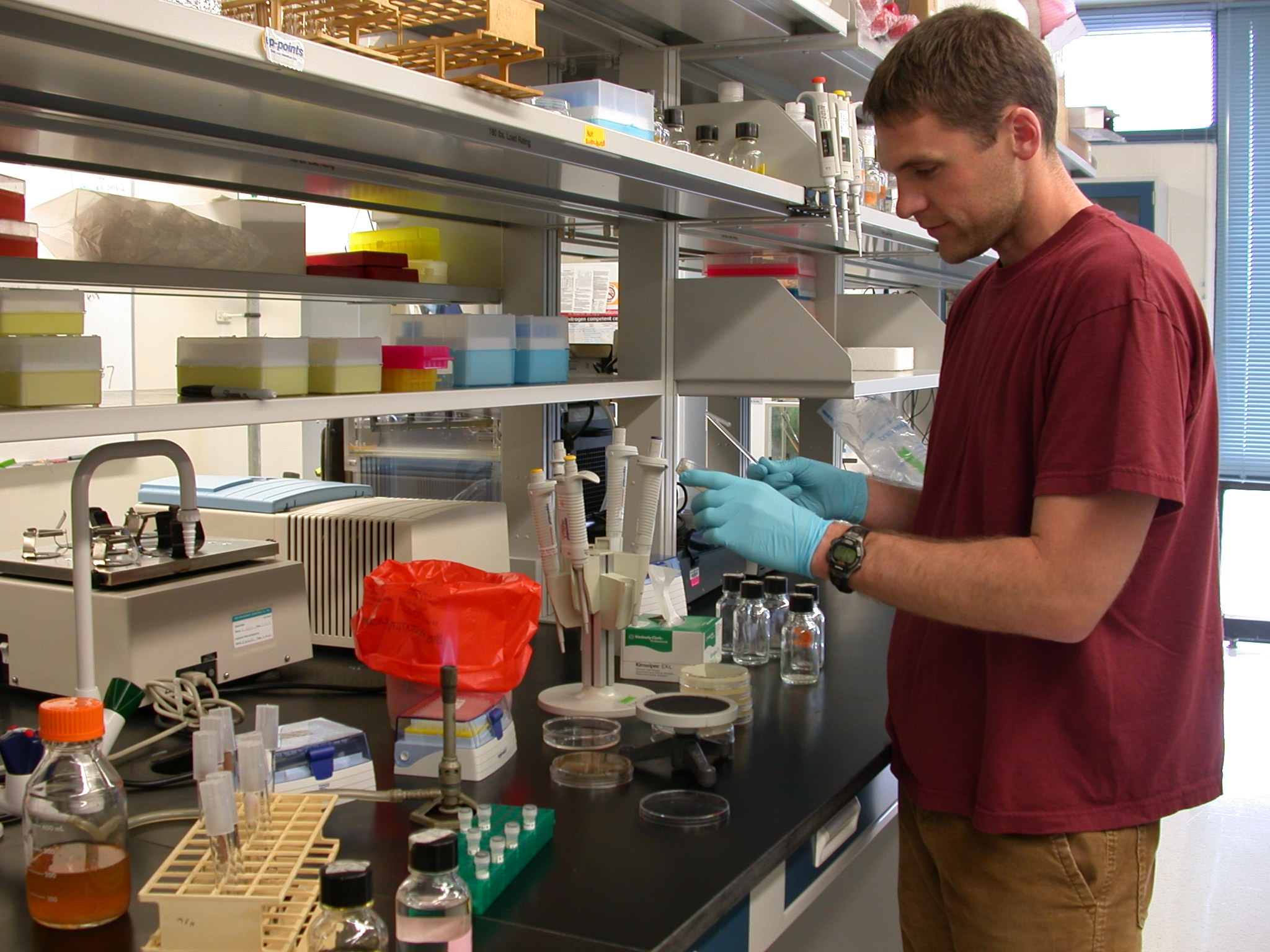
NanoEarth facilities at Kelly Hall includes a 3,300 sq. ft. laboratory with extensive nanoparticle synthesis facilities and a wide array of benchtop analytical instruments and an additional 3,000 sq ft. of meeting and office space for staff and visitors.
The laboratory is equipped to help users prepare environmental samples for analysis and to synthesize many types of nanomaterials that are also naturally occurring, so that they can be studied and better understood in the laboratory.
Our facilities are also equipped to produce many types of manufactured nanomaterials that can be inadvertently released into the environment, or used as an active agent in environmental remediation, or used in a nanomaterial-enabled detection device.
In all of these cases, laboratory production of these nanomaterials in carefully controlled experiments have a much higher degree of quality control (in terms of precise compositions, and more uniform sizes and shapes) than equivalent commercially available nanomaterials.
Instrumentation and Capabilities
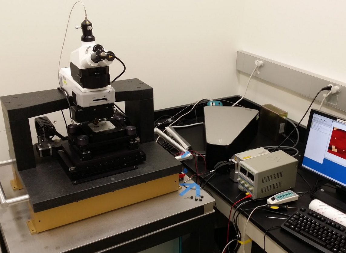
Wiitec Raman Confocal Atomic Force Microscope
The WITec alpha500 combines Confocal Raman Microscopy for 3-D Chemical Imaging and Atomic Force Microscopy for high resolution structural imaging in an automated system. It not only allows high resolution surface topography imaging with AFM at many different sample positions but also Raman Imaging, large area Raman scans and multi-point spectra acquisition at a user-defined number of measurement points. The Raman and AFM images of the same sample positions can then be matched and linked together for a comprehensive understanding of the sample's properties. Two lasers (633 nm and 785 nm) and multiple commonly used objectives including water immersion (63x) and oil immersion (100x) objectives for Raman measurements. Three measuring modes for AFM (contact mode, AC-mode and pulsed forced mode).
Veeco Multimode Atomic Force Microscope (AFM)
The Nanoscope IIIa Multimode AFM can be operated in both liquid and air environments. The image analysis and presentation software contains powerful algorithms for the measurement and presentation of research results including: cross sectional analysis, roughness measurement, grain size analysis, depth analysis, power spectral density, histogram analysis, bearing measurement and fractal analysis.
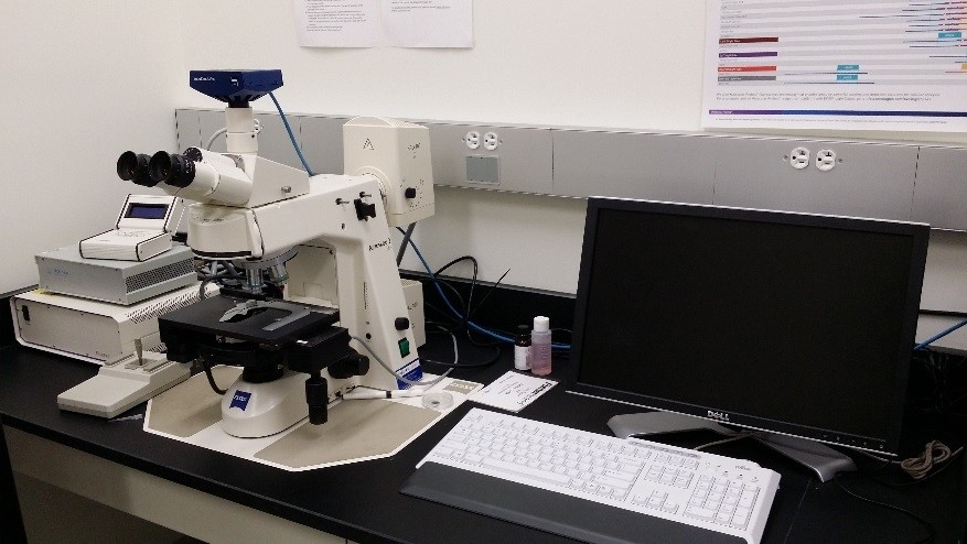
Zeiss Flourescence Microscope
With attached FluoArc, user can not only get the bright field image of a specimen, but also detect, localize and quantitate the amount of fluorescence in a specimen.
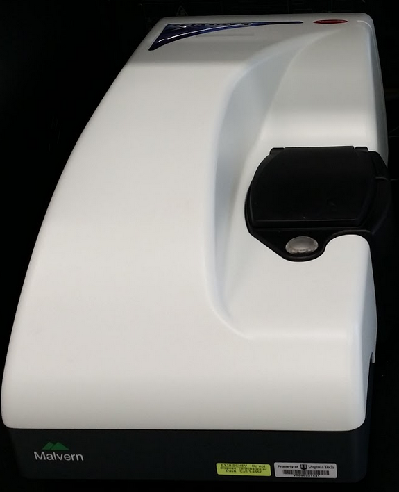
Dynamic Light Scattering (DLS)
Malvern Zetasizer Nano ZS can measure particle size/ size distribution and zeta potential in either aqueous or organic solvent.
High Performance Liquid Chromatograph (HPLC)
The Hewlet Packard HPLC is used to separate, identify, and quantify each component in a mixture.
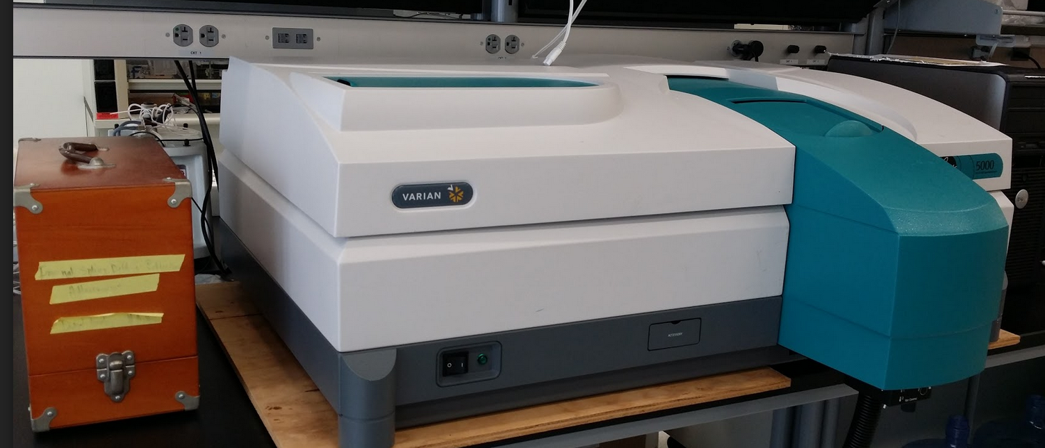
Cary 5000 UV-Vis-NIR Sepctrometer
The Cary 5000 is a high performance UV-Vis-NIR spectrophotometer with super photometric detection in the 175-3300 nm range. The system can perform absorption, transmission and diffusion reflectance scans depending on the sample status. A multi-sample holder with temperature controlling is convenient for any dynamic analysis.
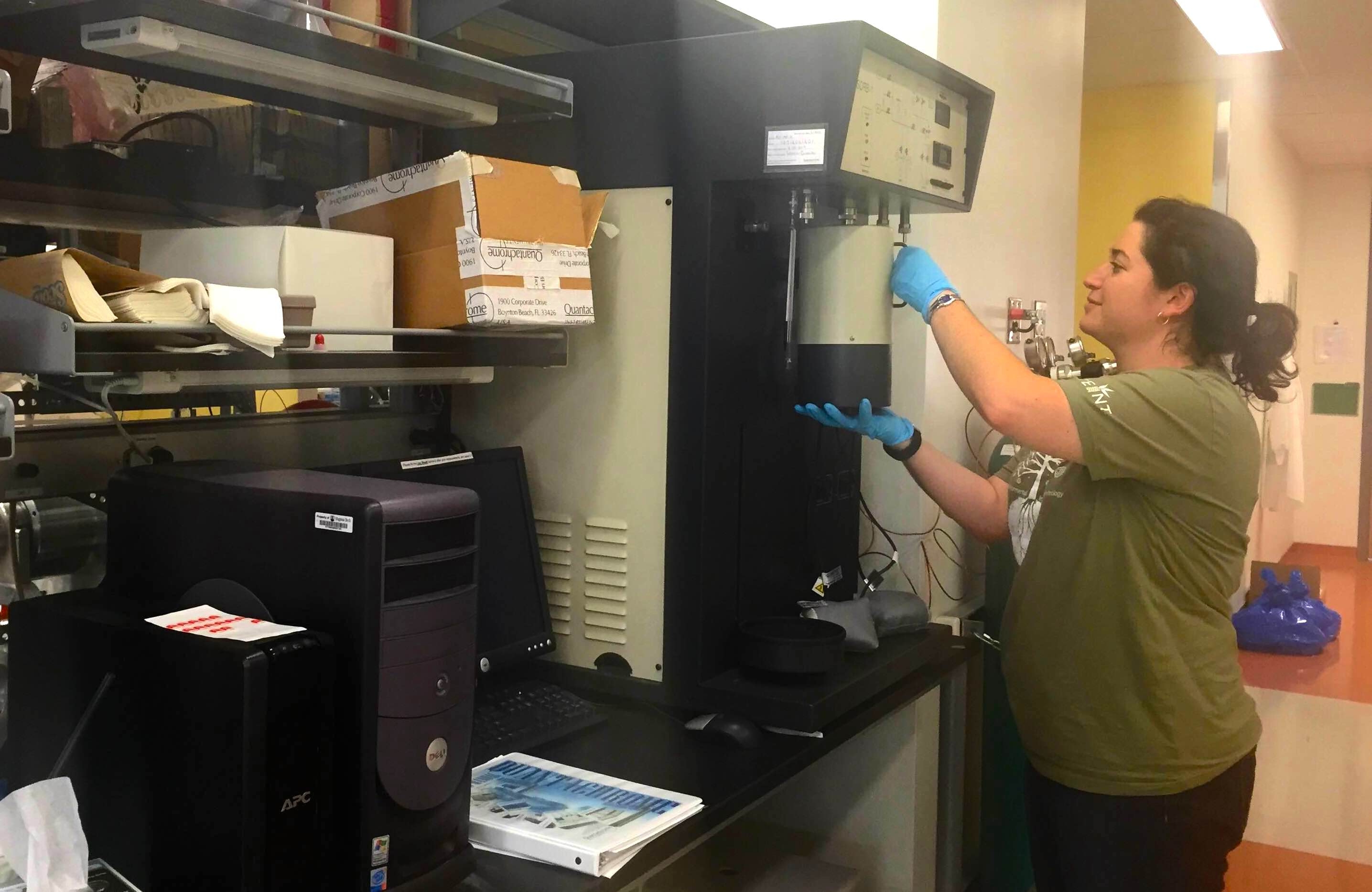
Quantachrome AUTOSORB-1
Quantachrome AS-1 has the capability of measuring adsorbed or desorbed volumes of nitrogen and then calculating BET surface area (single and/or multipoint), Langmuir surface area, adsorption and/or desorption isotherms, pore size and surface area distributions, micropore volume and surface area using an extensive set of built-in data reduction procedures.
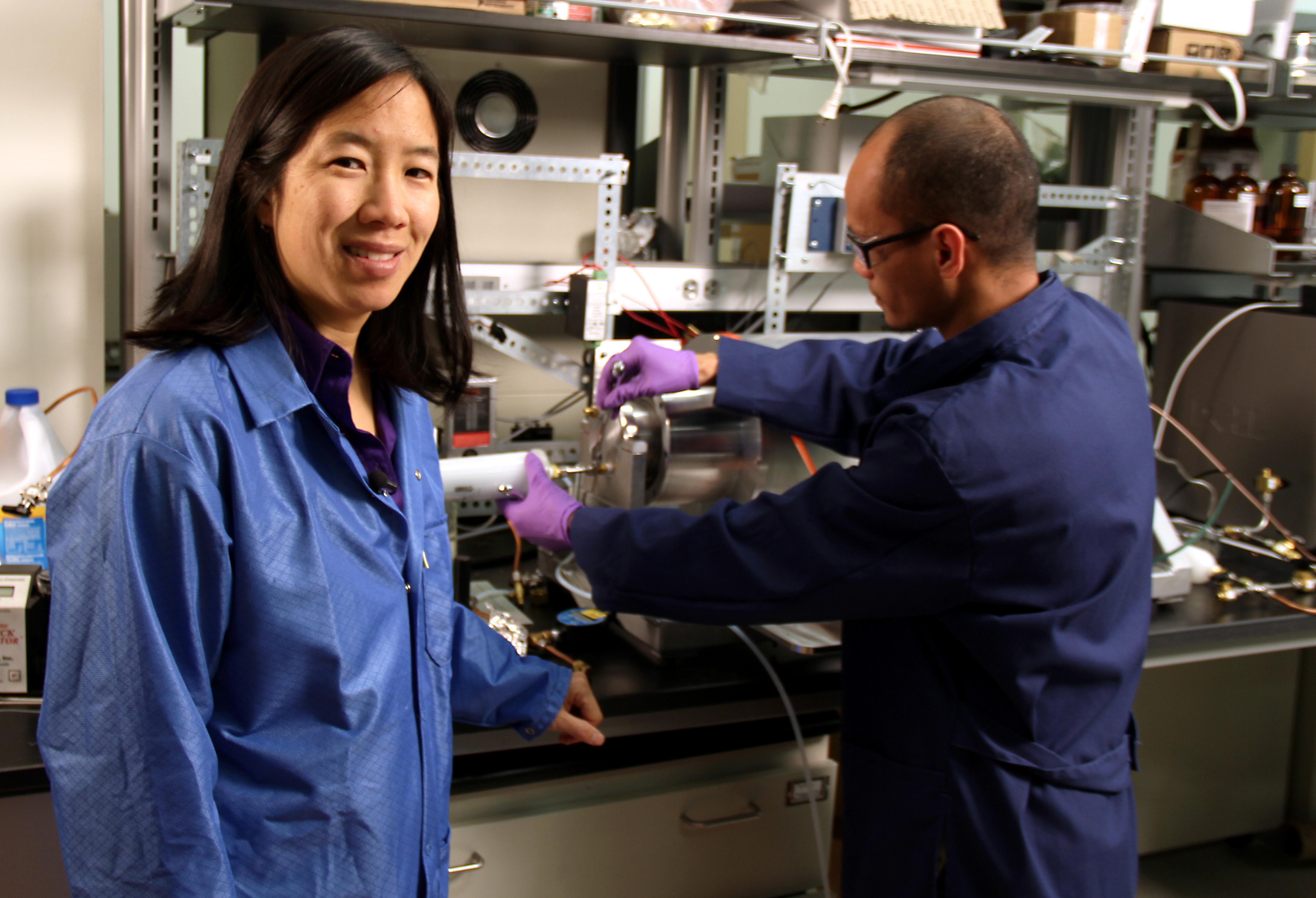
A 6 m3 polytetrafluoroethylene (PTFE, Teflon ®) chamber for aerosol chemistry experiments.
TSI 3936NL Scanning Mobility Particle Sizer (SMPS)
Capable of measuring high-resolution size distribution of aerosols spanning 4 - 700nm.
TSI 3321 Aerodynamic Particle Sizer (APS) Spectrometer
Provides high-resolution, real-time aerodynamic measurements of particles from 0.5 to 20 μm. Includes an additional diluter for high concentrations.
TSI 9306 Aerotrak Optical Particle Counter
Handheld Particle Counter for aerosols ranging from 0.3 to 25 μm.
TSI DustTrak 8520 PM Monitor
Aerosol photometer for real-time measurement of PM1, PM2.5, or PM10.
EcoChem Analytics DC 2000 CE Diffusion Charger
For real-time measurement of aerosol surface area.
EchoChem Analytics PAS 2000
Photoionization aerosol sensor for particulate PAH.
Sioutas impactors
Personal cascade impactors in five size ranges including > 2.5, 1.0 to 2.5, 0.50 to 1.0, 0.25 to 0.50, and < 0.25 μm. Samples are collected onto 25-mm PTFE filters.
California Measurements MPS-3 Microanalysis Particle Sampler
A 3-stage cascade impactor with particle size cut-points down to 0.05 μm. Samples are collected onto SEM stubs or TEM grids.
Multiple themophoretic precipitators made in-house.
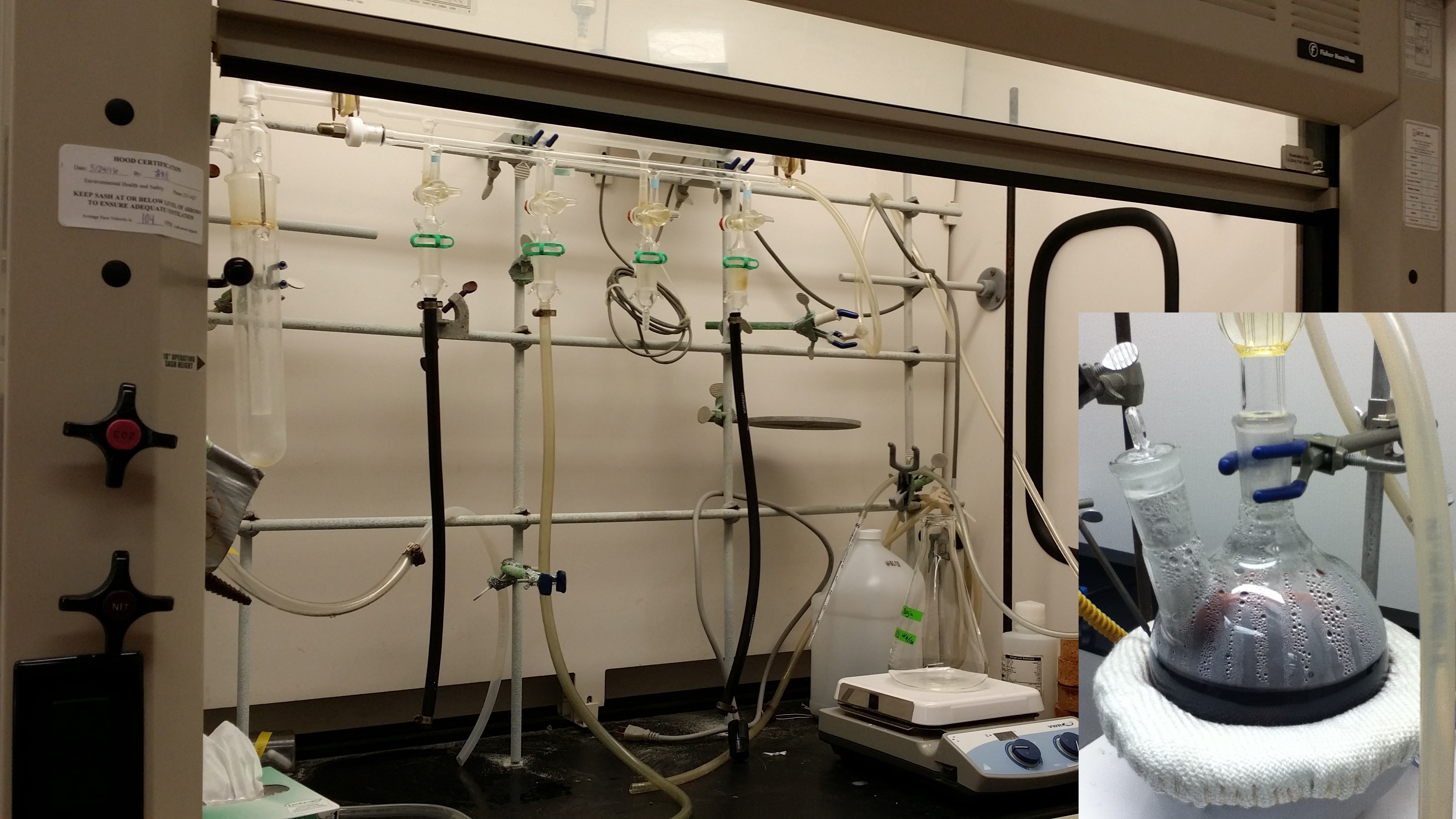
Examples of nanomaterials that have been synthesized in our facilities include:
Gold nanomaterials:
- Medium Temperature, Gold nanoparticles (AuNP)
- Elevated Temperature Seed-Mediated AuNP Growth
- Room Temperature Seed-Mediated AuNP Growth
- Gold Nanorods (AuNR)
- Pure AuNP multimers
- AuNP Surface Functionalization Introduction, and Polymer Functionalization
- Nanocellulose/Gold Nanoparticle Nanocomposites
Silver Nanoparticle (AgNP)
Medium Temperature, Organic Solvent-Based Chalcogenides:
- Galena (PbS) nanocrystals.
- Sphalerite (ZnS) nanocrystals.
Room Temperature, Abiotic Fe-/Mn-(Oxyhydr)oxide Nanoparticles:
- Hematite (Fe2O3) and Magnetite (Fe3O4) nanoparticles.
- Manganese Oxide (Mn3O4) nanowires.
- Green Rust (a ferric/ferrous oxyhydroxide sulfate/carbonate hydrate).
Room Temperature, Bacteria-Directed Synthesis of Metal-Sulfide Nanoparticles:
- Sphalerite [(Zn,Fe)S]
- Iron Sulfides (FeSx)
Various other instruments for producing/detecting/sampling aerosol gases and particles.
Ultrapure water filtration system (Barnstead Nanopure Life Science with TOC monitor).
Pellicon cross-flow ultrafiltration (CFUF) system (Millipore) for water sample separation and concentration.
5 chemical fume hoods and 1 biological laminar flow hood.
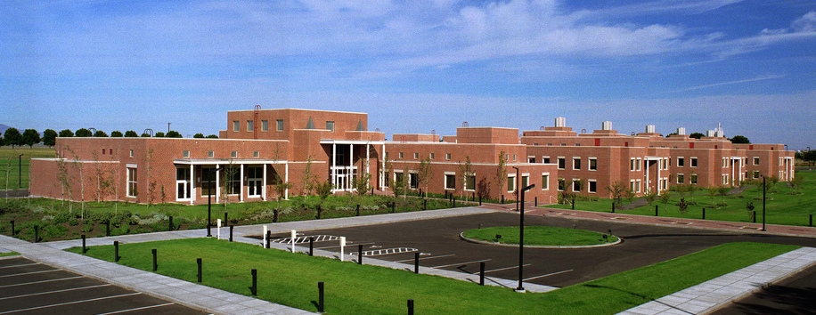
NanoEarth has established a partnership with EMSL at PNNL to direct users whose specific needs may be better met by their state of the art facilities.
EMSL’s mission as a national scientific user facility is to lead molecular-level discoveries for the Department of Energy and its Office of Biological and Environmental Research that translate to predictive understanding and accelerated solutions for national energy and environmental challenges.
EMSL's unparalleled collection of experts, advanced instrumentation and state-of-the-art facilities can help scientists gain a predictive understanding of the molecular-to-mesoscale processes in climate, biological, environmental and energy systems. These advancements are critical to development of sustainable solutions to the nation's energy and environmental challenges.
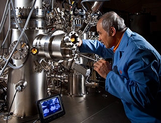
The scientific capabilities at EMSL include:
- A variety of NMR-based instruments, including next-generation sequencing platforms,
- Atomic force, confocal, fluorescence, and FTIR microscopes,
- Cryo and environmental transmission electron microscopes (TEM),
- Scanning electron microscopes (SEM),
- Environmental focused ion beam (FIB), Ion Etching,
- Atom probe tomography, electron microprobe, electron spectrometers,
high resolution EELS and XPS, - X-ray computed tomography (XCT),
- Multiple mass spectrometers,
- A 3.4 petaflop computing system
and much more.
The Environmental Nanoscience and Analytics Laboratory is an interdisciplinary research team focusing on understanding the environmental exposure, interactions, fate, effects, and applications of engineered nanomaterials.
Applications
- Elemental analysis: Elemental analysis (all elements, all isotopes at all times) of environmental, geological, and other types of samples
- Single particle analysis: Quantitative analysis of all elements and all isotopes in nanoparticles on a particle-per-particle basis
- Continuous fractionation of nanomaterials: High resolution fractionation and size distribution analysis of polydispersed nanomaterial suspensions. Elemental composition as a function of particle size when coupled with ICP-TOF-MS.
- Elemental imaging: High speed, high resolution, full mass spectrum imaging of biological, geological and other samples Sample preparation We also support sample preparation such as microwave assisted acid digestion for total elemental analysis and nanoparticle extraction for SP-ICP-TOF-MS and AF4-ICP-MS analysis. We can also discuss and provide sample preparation protocols to interested users.
Contact
Dr. Mohammed Baalousha
Associate Professor
ENHS/ASPH (PHRC 501C)
mbaalous@mailbox.sc.edu
Dr. JingJing Wang
Lab Manager
ENHS/ASPH (PHRC 413)
jingwang@mailbox.sc.edu
Instrumentation and Capabilities
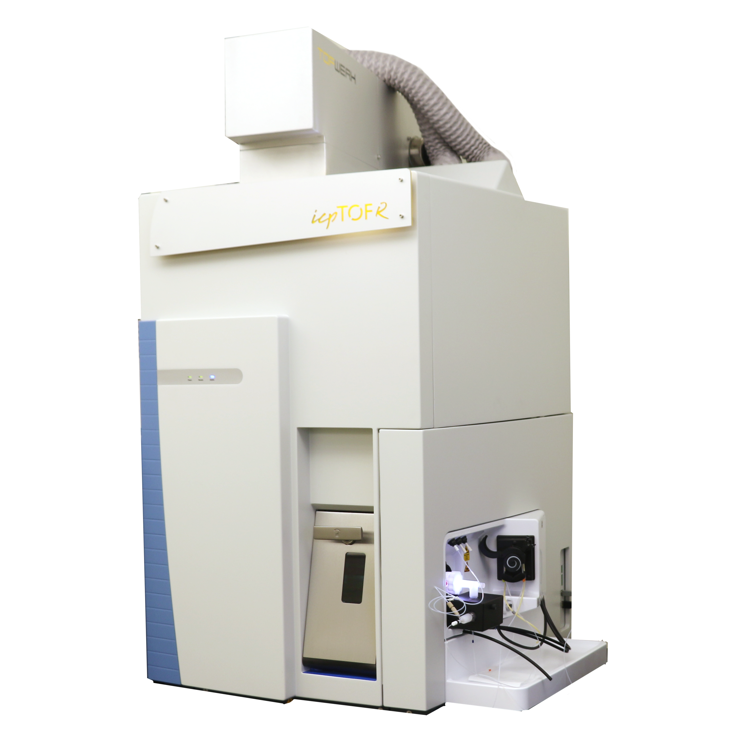
Inductively coupled plasma-time of flight mass spectrometer (ICP-TOF-MS) combines a Thermo Scientific iCAP RQ to a TOFWERK TOF mass analyzer. The ICP-TOF-MS measures all elements, all isotopes, all the time in transient signals such as single particle, single cell, and laser ablation imaging at mass resolving power > 3000.
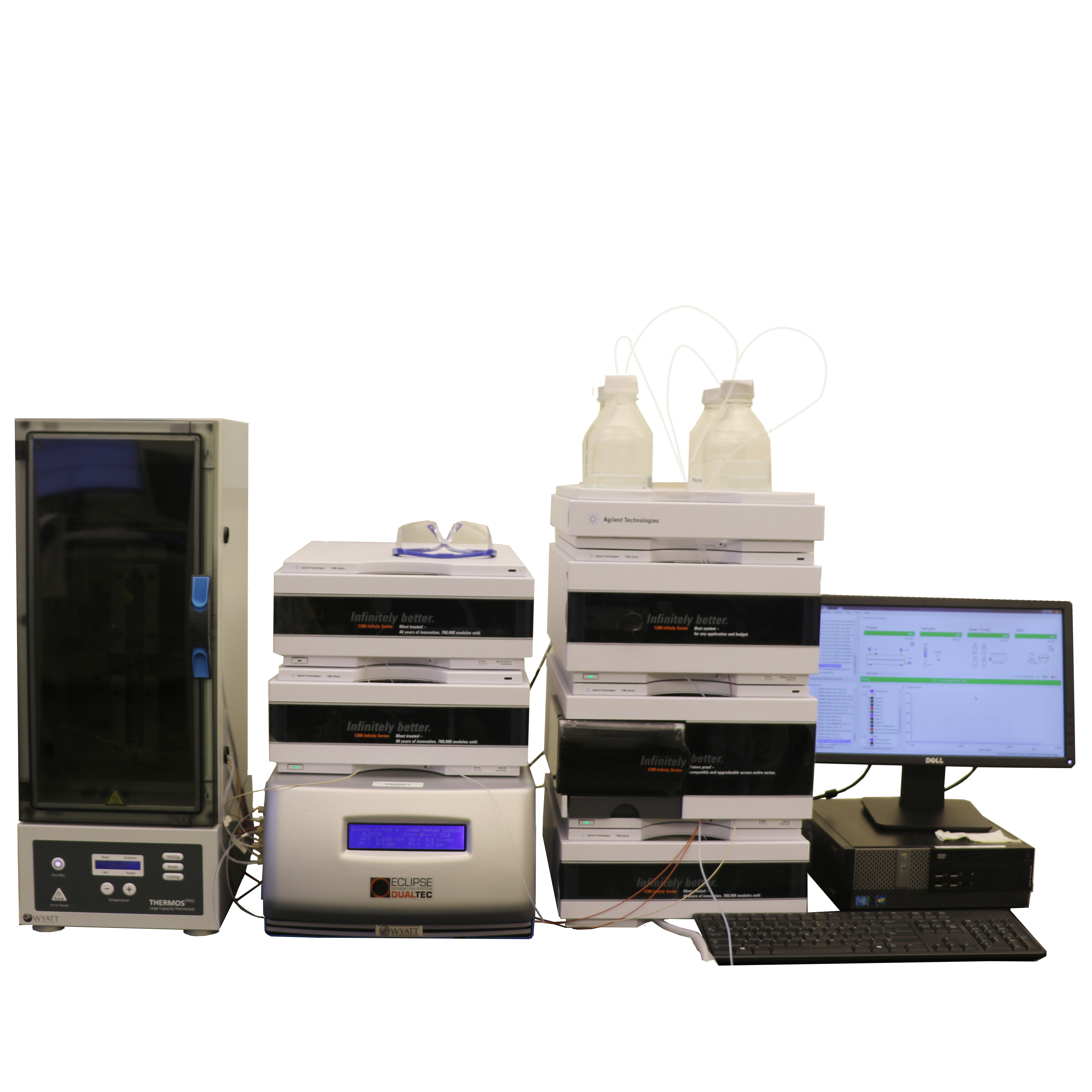
The asymmetrical flow-field flow fractionation (AF4) allows high resolution fractionation of nanomaterials and elemental composition analysis when coupled with ICP-TOF-MS.
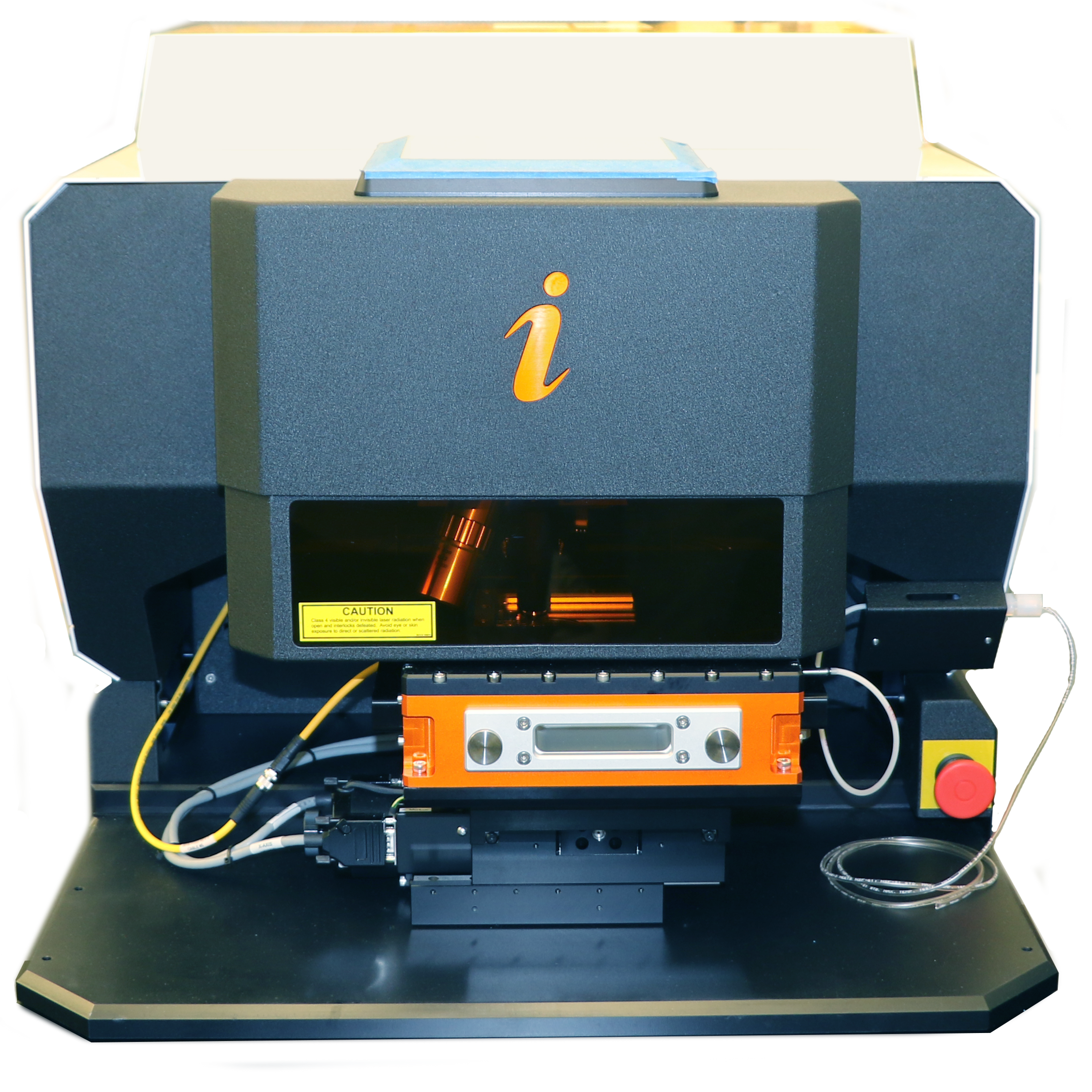
Image Bio Laser Ablation System enables high resolution, high-speed, bioimaging by Laser Ablation-ICP-TOF-MS.
ENAL also supports sample preparation such as microwave assisted acid digestion for total elemental analysis and nanoparticle extraction for SP-ICP-TOF-MS and AF4-ICP-MS analysis. We can also discuss and provide sample preparation protocols to interested users.
Dr. Baalousha and his team can also support data analysis, interpretation, and contribute to writing the data for scientific collaboration. In particular, the SP-ICP-TOF-MS generates large and complex data sets comprising multi-element composition of thousands of particles. We developed and continue to develop unsupervised machine learning approaches to identify classes of nanoparticles based on their elemental composition.


