

Focused Ion Beam
APPLICATIONS
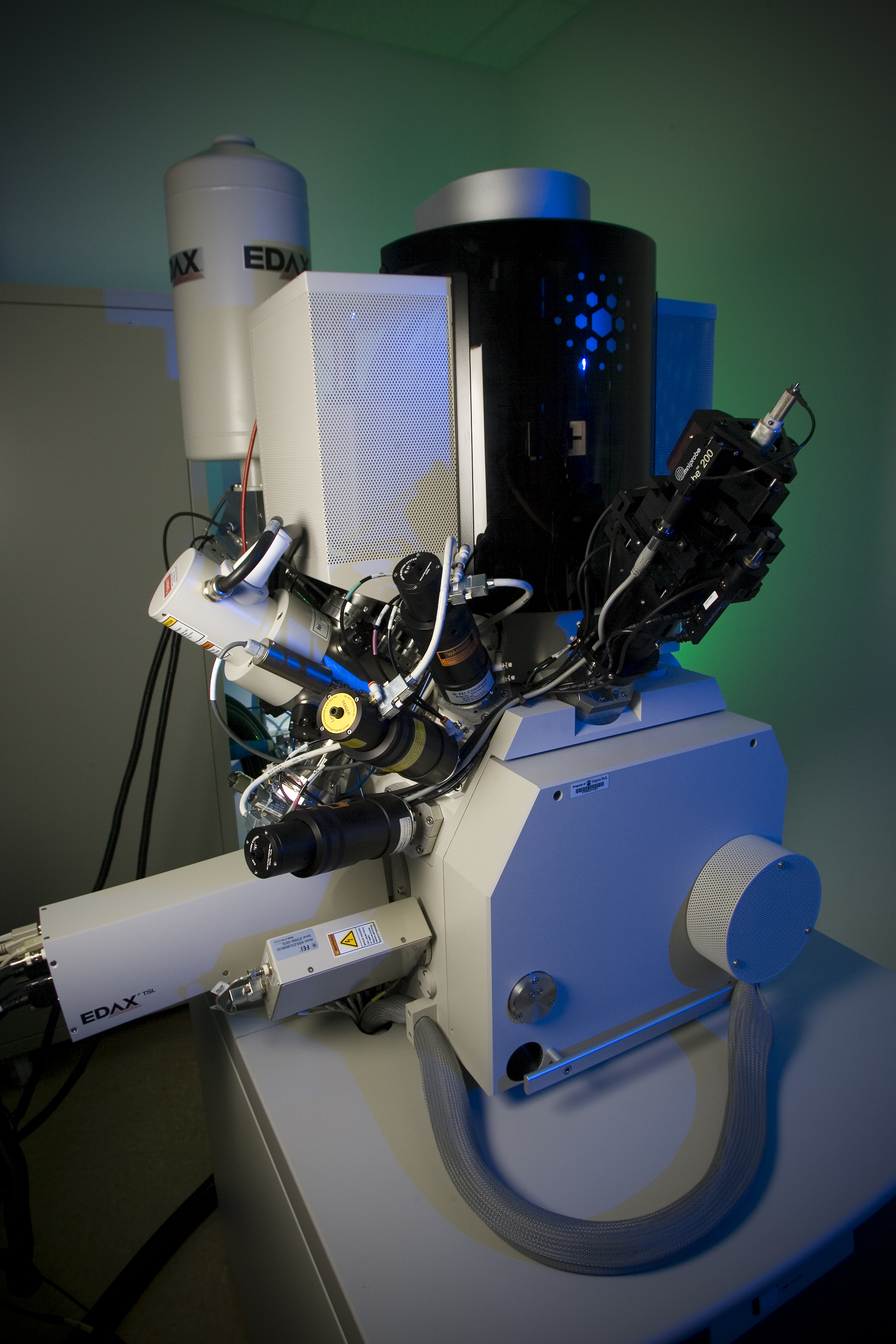
Dual beam FIB-SEM is a workstation that combines a scanning electron microscope (SEM) and a focused ion beam (FIB); it is heavily used in semiconductor industry, materials science, and increasingly in geoscience and biological field for site-specific analysis. SEM delivers high resolution images, while FIB is mostly used to add and subtract micro volumes of material in a controlled manner, like a nanomachining device. Coupled to an Energy Dispersive X-ray Spectroscopy (EDS) detector and Electron Backscattered diffraction (EBSD) camera, dual beam FIB-SEM provides elemental identification and crystallographic properties from the cross-section of the sample. With serial slicing technique, structural, chemical and crystallographic information can be revealed in three dimensions.
- High resolution cross-section images
- Site-specific liftout preparation for TEM analysis
- Grain contrast imaging
- Circuit modification
- Serial slicing and 3D reconstruction
LIMITATIONS
- Vacuum compatibility required
- Sample size restriction
- Ion implantation
- Ion beam damage
Schindler, M., Michel, S., Batcheldor, D., & Hochella Jr., M.F. (2019). A nanoscale study of the formation of Fe-(hydr)oxides in a volcanic regolith: Implications for the understanding of soil forming processes on Earth and Mars. Geochimica et Cosmochimica Acta, 264, 43–66.
Invalid iframe embed code.