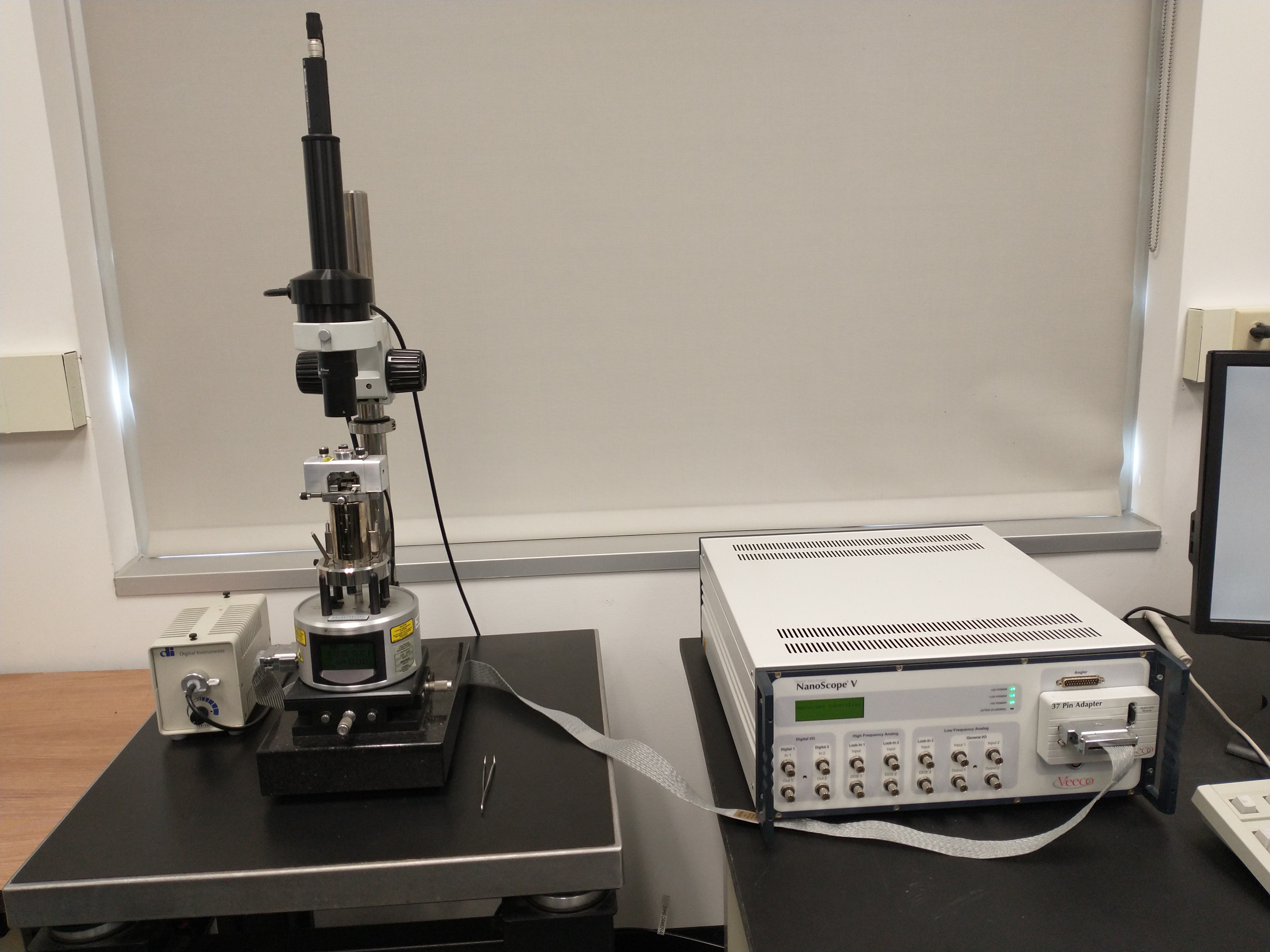
Atomic Force Microscopy

Atomic force microscopy (AFM) is a very-high-resolution type of scanning probe microscopy (SPM), with demonstrated resolution on the order of fractions of a nanometer, more than 1000 times better than the optical diffraction-limit.
APPLICATIONS
- Three-dimensional surface topographic imaging, including surface roughness, grain size, step height, and pitch
- Nanoscale imaging of polymer structure and morphology
- 2D material property measurements
LIMITATIONS
- Scan range limits: 125 µm laterally (xy) and 5 µm vertically in z-direction
- Potential problems with extremely rough or oddly shaped samples
- Limited sample size: < 15 mm diameter, 5 mm thick
Liu, C., Leng, W., & Vikesland, P. J. (2018). Controlled Evaluation of the Impacts of Surface Coating on Silver Nanoparticle Dissolution Rates, ES&T, 52(5), 2726-2734.
The safety issues with nanoparticles are continuously being tested since their potential dangers for the environment and human health. Dissolution of nanoparticles is an important process that alters their properties and is also a critical step in determining safety of nanoparticles. Distinguishing the toxicity of nanoparticles in particle form relative to that of the released ions is a critical but challenging research topic. Studying nanoparticle dissolution as illustrated herein can help in the current move toward safer design and application of nanoparticles. Environmental risk assessments of engineered nanoparticles require characterization of nanoparticles and quantitative analytical methods to determine environmental concentrations. The effect and exposure of engineered nanoparticles are vital for the development of related products. In this project, the uniform arrays of nanoparticles enable the controlled evaluation of nanoparticle dissolution in the absence of aggregation. We tracked the height changes of AgNPs fabricated on glass slides using Atomic Force Microscope (AFM). The controlled evaluation of height changes were obtained by AFM measurements. The effects of surface coating on nanoparticle dissolution were illustrated in the absence of aggregation. Information obtained from this study demonstrated different coating agents exhibit various effects on the dissolution process. Specifically, the PEG coating prevents the AgNPs from dissolving in 2 weeks which lead almost no Ag+ released to the solution. For BSA coated samples, slight changes were observed for the shape and height. The results of this work will aid efforts to engineer environmentally benign nanomaterials and to predict the environmental impacts of coated NPs.
Figure 1 (a) Schematic of AgNP array production and coating process, (b) AFM image of original AgNPs, and (c) AgNP height distribution as measured by AFM. The mean height of 585 particles was 47.1 with a standard deviation of 1.5 nm.


