2023 Plenty of Beauty at the Bottom Image Contest
The Plenty of Beauty at the Bottom image competition was fierce this year with so many amazing images competing for the Virginia Tech slots in the NNCI contest! Below are the three entries that we have selected to move on to the NNCI competition (https://www.nnci.net/plenty-beauty-bottom).
Local Winners - Submitted to NNCI Competition
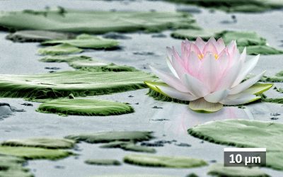
Submission Category: Most Stunning
Title: Biochar Beach: Surfing the Nano Seas
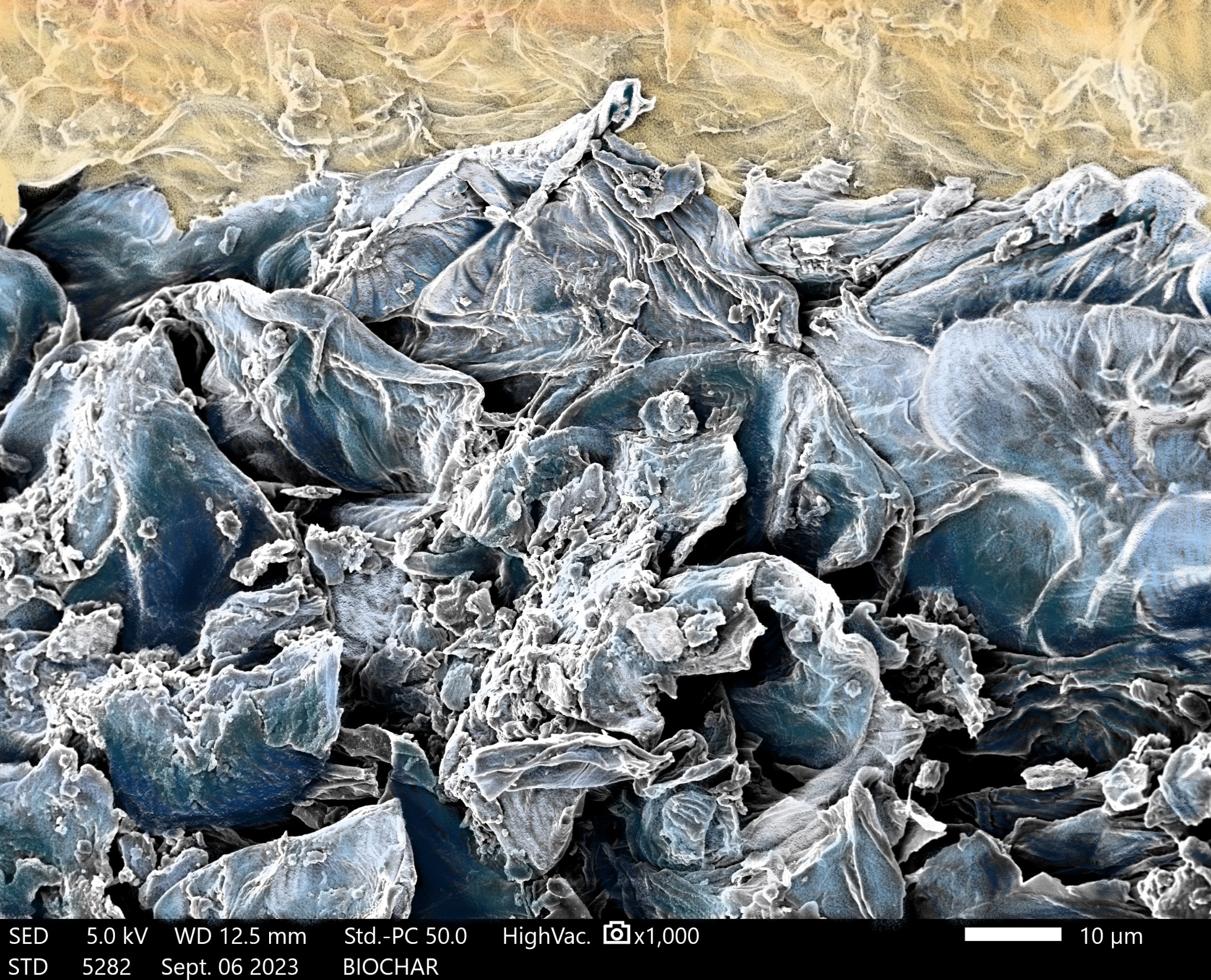
Artists: Yilin Li, Ph.D. Candidate, Department of Food Science and Technology, College of Agriculture and Life Sciences, Virginia Tech
Tool: JEOL IT-500HR Scanning Electron Microscope
Short Description: The overall goal of the research is to develop a cost-effective filtration system that utilizes biochar produced from food waste to effectively reduce pathogens by adsorption. SEM images were used to assess the porosity and surface morphology of biochar derived from pecan shells carbonized at a temperature of 300 ºC. The images reveal a sheet-like structure, indicating potential for high surface area. SEM images enable the researchers to have a better understanding of the surface characteristics of biochar produced at a specific temperature and provide insight into its potential adsorption and filtration efficiency.
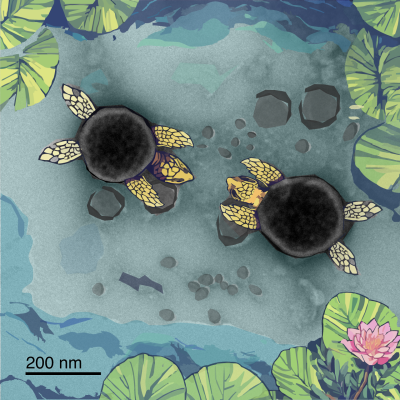
Submission Category: Most Unique Capability
Title: Nanoplastic Volcano Ball!
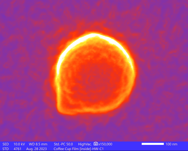
Artists: Bipin D. Lade, Ph.D., Postdoctoral Associate, NanoEarth, Virginia Tech and F. Marc Michel, Ph.D., Associate Professor, Department of Geosciences, College of Science, Virginia Tech; Division Leader of Nanoscience, Academy of Integrated Science, College of Science, Virginia Tech; and Deputy Director, NanoEarth, Virginia Tech
Tool: JEOL IT-500HR Scanning Electron Microscope
Short Description: The SEM image shows a single nanoplastic particle located on the surface of the inner lining of an unused single-use beverage cup. Micro-sized and nanosized plastic particles are released from these liners when exposed to water during use. Our laboratory is actively developing sampling protocols for nanoscale characterization and conducting research on nanoplastic synthesis and aggregation using hydrophobic fluids. Our primary focus lies in the characterization of nanoscale materials, and we utilize a range of analytical instruments, including transmission electron microscopy (TEM), scanning transmission electron microscopy (STEM), Raman spectroscopy, atomic force microscopy (AFM), dynamic light scattering (DLS), and UV-VIS spectroscopy.
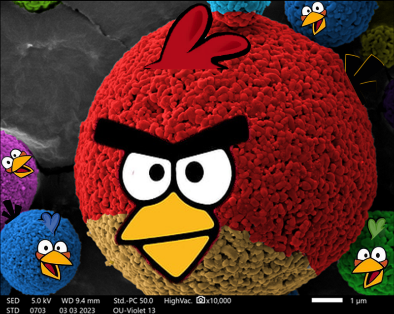
Submission Category: Most Whimsical
Title: Micro-Nano Angry Birds!

Artists: Bipin D. Lade, Ph.D., Postdoctoral Associate, NanoEarth, Virginia Tech and Ethan Franklin, Undergraduate Student, Division of Nanoscience, Academy of Integrated Science, College of Science, Virginia Tech
Tool: JEOL IT-500HR Scanning Electron Microscope
Short Description: The SEM image shows spherical microparticles composed of hematite nanoparticles in a pigment created from materials collected from acid mine waste drainage. Our laboratory specializes in nanoparticle synthesis and materials characterization using analytical instruments, including TEM, STEM, Raman, AFM, DLS, and XRD. We explore the formation of the smallest minerals, known as nanoparticles (NPs), their interactions with their surroundings, and their transformations over time in complex natural and environmental systems.
Congratulations to Yilin, Bipin, Marc, and Ethan! Good luck in the full competition!
Honorable Mentions
We also have a couple of fun honorable mentions that we loved so much we had to share.
Submission Category: Most Stunning
Title: Shattered World
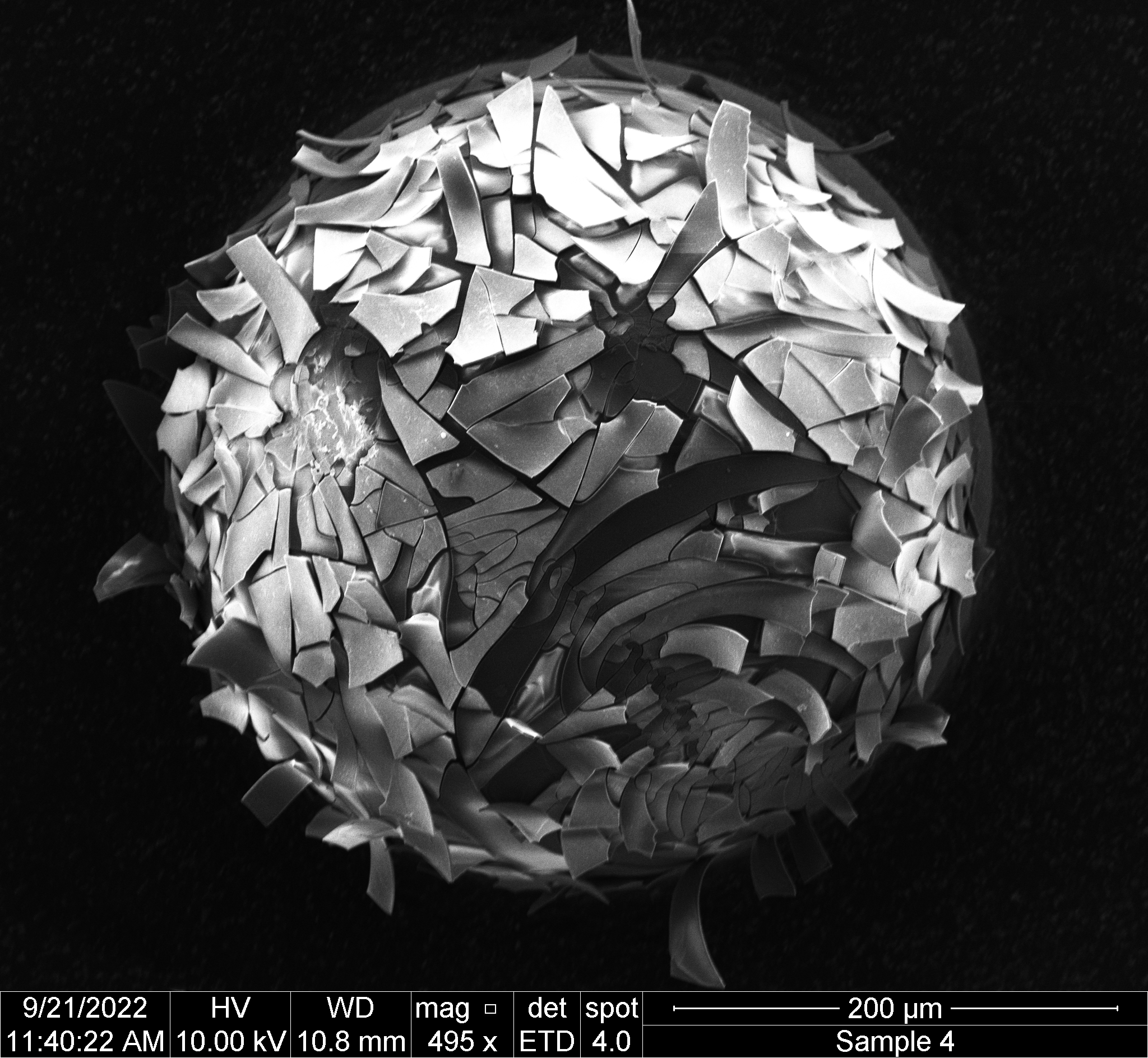
Artists: Elliott Kunkel, Ph.D Candidate, Department of Chemical Engineering, College of Engineering, Virginia Tech and Hans Robinson, Ph.D., Associate Professor, Department of Physics, College of Science, Virginia Tech
Tool: FEI Quanta 600 FEG Scanning Electron Microscope
Short Description: In the captured image using the FEI Quanta 600 FEG, a shattered barium titanate microsphere with a coating of poly(dopamine) set against a backdrop of carbon tape is observed. It is not uncommon to find air pockets encapsulated within these barium titanate spheres, a phenomenon attributed to their fabrication process. We hypothesize that during the vacuum stage of sample preparation, the sphere succumbed to implosion, culminating in the distinct fractured-world pattern.
Our laboratory specializes in the research of patchy particles, emphasizing their potential for self-assembly. We are actively investigating various catecholamine coatings to analyze the impact of both composition and metal salt concentration on the photo-induced deposition of metal ions on the surface.
Submission Category: Most Whimsical
Title: Sandpipers on a Carbon Nanotube Shore
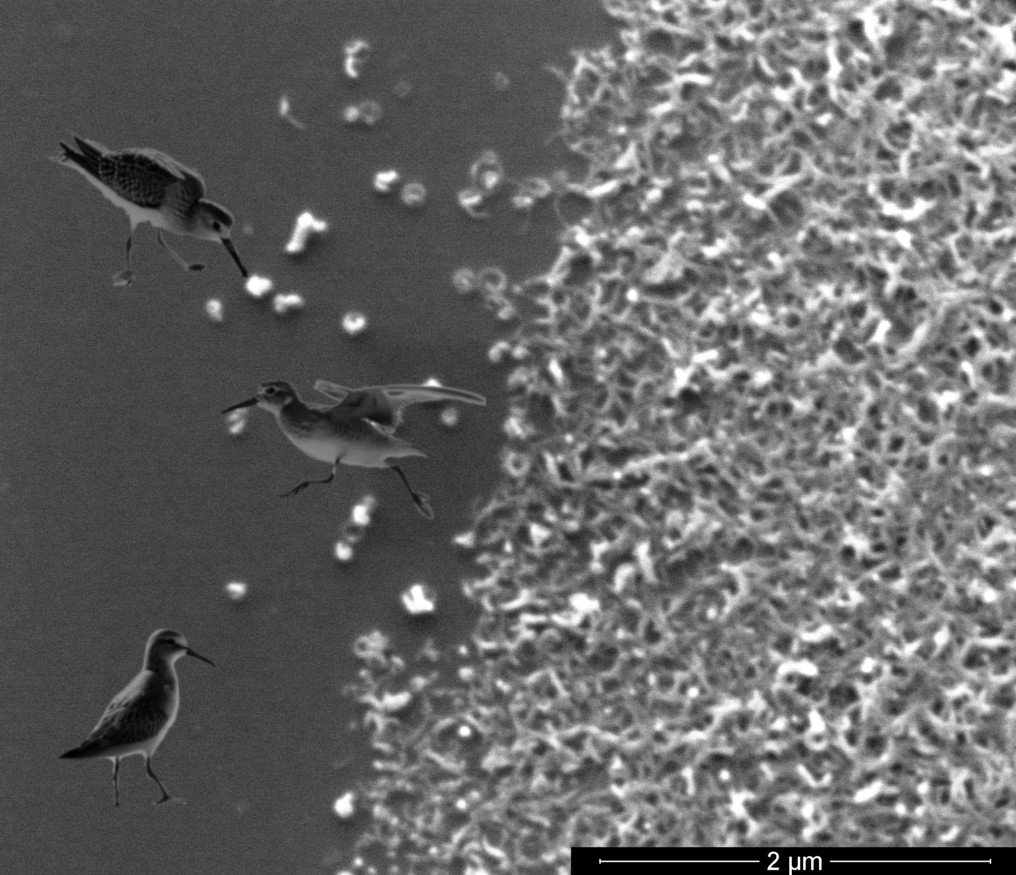
Artists: W. Cary Hill, Ph.D., Senior Research Scientist, ITA International, LLC
Tool: FEI Quanta 600 FEG Scanning Electron Microscope
Short Description: Commercially acquired multiwall carbon nanotubes were distributed on a silicon wafer as part of an effort to validate claimed morphological properties. The edge of the carbon nanotube deposit has the appearance of a seashore; microscale sandpipers are inserted to helpfully collect stray nanotubes escaping from the tangled mat.